“DNA neither cares nor knows. DNA just is. And we dance to its music.” – Richard Dawkins, evolutionary biologist and zoologist.
You may have been told that your DNA makes you what you are. That is indeed true, since DNA is the master guide telling your cells (which work together to make you up) how to do their thing and survive. But how does a clump of nucleic acid define what we are? How does it magically know what to do, when to do it and why it has to do it? This series of articles covering the principles of genetics hopes to answer those questions (also in accordance with the H2 Biology curriculum). The genomics mega-chapter will talk about all things genetics and nucleic acids, from what the genome is, how nucleic acid codes get translated into functional physiological phenomena and how these phenomena affect us as living beings. That’s a lot to cover, so this article will first cover some foundational concepts, which we will regularly visit in upcoming chapters.
The genome refers to the entire set of genetic material in an organism, or the entirety of an organism’s hereditary information. It is encoded as either DNA (as in eukaryotes and prokaryotes) or as RNA (as in the case of many viruses). The genome of an organism is replicated before cellular replication, and includes both coding and non-coding sequences. Such genes can be categorized into structural genes (protein and RNA-encoding genes), functional sequences (regulatory sequences such as initiation/promoter sites etc.) and non-functional sequences (Introns and repetitive sequences, heavily involved in regulation of gene expression).The expression of genes within the genome produces proteins that keep physiological and cellular processes running.
But before you go on reading… You might want to download a pdf copy of this article as it is quite long!
Click the ‘Download’ button, enter your email, and the pdf file will be delivered to your inbox! (Remember to check spam!)

The cell cycle is the circle of life for a cell! Our cells undergo rounds of cellular division to produce more cells for growth and to replace dying or damaged cells. The cell cycle refers to a series of events that lead to cellular division, and can be separated into 3 stages: G0, Interphase (G1, S and G2) and mitotic (M) phase. Before proceeding from one stage to another, cell cycle checkpoints ensure that appropriate and sufficient progress has been made. Checkpoints help to prevent errors and ensure accurate cell division. Loss of checkpoint function can lead to genomic instability, which results in mutations (to be covered later!). Mutations in checkpoint proteins are a common hallmark in many types of cancer!
Most of the time, the cell is not actively dividing and remains in the resting G0 state, where it continues with its regular cellular activities.
When stimulated to undergo cellular division (such as a growth signal), the cell enters the interphase. During interphase, the cell undergoes several processes in preparation for mitosis such as DNA replication, cellular growth, synthesis of macromolecules and cell organelles. This stage of the cell cycle usually lasts 23 hours.
In the G1 phase, the cell grows in size and synthesizes the organelles and macromolecules it will need. Synthesis of DNA replication machinery will be upregulated in preparation for the upcoming S-phase. As cell division requires a lot of energy, the cell accumulates a lot of energy over the course of the G1 phase.The cell undergoes DNA replication during the S phase, also known as the synthesis phase. The centrosome also undergoes duplication during the S-phase in preparation to form the mitotic spindle during mitosis. By the end of the S phase, the cell would have double the amount of DNA in its nucleus.
The G2 phase is the second gap phase following the S phase. During this phase, the cell continues to grow and synthesise organelles and proteins. The final preparations for mitosis are completed during this phase.
Mitosis takes place during the M phase. The cell undergoes the processes of prophase, metaphase, anaphase and telophase, which are the stages of mitosis (to be covered later). After mitosis, the cell splits into its 2 daughter cells over cytokinesis. The daughter cells enter G0 awaiting for the next signal for cellular division.
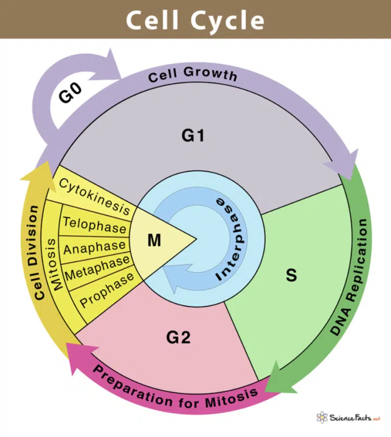
Fig 1. A diagram describing the cell cycle.
Non-dividing cells are in the G0 phase, and can enter G1 when stimulated to divide. G1 is the longest phase of the cell cycle, during which the cell grows in size and synthesizes new organelles and macromolecules. This is followed by the S phase, where DNA replication occurs. The G2 phase takes place after S and the cell makes its final preparations for mitosis. Finally, mitosis occurs in the M phase and the cell divides into two daughter cells after cytokinesis.
Nucleotides are the building blocks of nucleic acids like DNA and RNA, and generally consist of a pentose sugar, with a phosphate group attached to its 5’ carbon and a nitrogenous base attached to the 1’ carbon. In RNA, the pentose sugar is ribose and has an additional hydroxyl group on the 2’ carbon. However in DNA, the pentose sugar is deoxyribose which lacks the 2’ hydroxyl group. This contributes to the stability of DNA as there is no additional hydroxyl group vulnerable to chemical reactions
In both DNA and RNA, there are four nitrogenous bases that form pairs with each other through hydrogen bonds. In DNA, the bases are adenine (A), thymine (T), guanosine (G) and cytosine (C), where A forms 2 hydrogen bonds with T while C forms 3 hydrogen bonds with G. This is known as complementary base pairing and A/T and C/G are known as base pairs. In RNA, the nitrogenous base thymine is replaced with uracil (U) that also forms 2 hydrogen bonds with A.
Complementary base pairing allows for nucleic acid sequences to be replicated easily, also allowing for storage and replication of genetic information. Many DNA and RNA-binding proteins recognize specific base-pairing patterns in order to bind to their target site. Even though base pairing is extremely accurate, mistakes do happen (rarely) and the wrong base gets paired up. This phenomenon is known as a mutation and results in changes to the genetic sequence. However, not all mistakes are catastrophic and some may even result in happy accidents that improve the survival of an organism. For reasons explained above, DNA is the material of choice for storing genetic information for higher organisms, due to its stability, replicability, information storage and potential for mutations. As genetic material, DNA exists as a double helix, with 2 long strands of polynucleotides running in opposite directions against each other (anti-parallel). Each nucleotide is connected to another nucleotide through a phosphodiester bond, where the 5’ phosphate group forms 2 ester bonds to link two nucleotides together. The direction of each strand can be deduced based on where the 5’ phosphate group of each strand is facing. Directionality will be extremely important, as DNA replication, transcription and RNA translation will involve reading the nucleic acid strand in various directions!
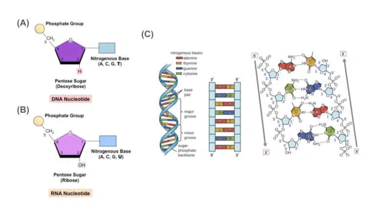
Fig 2. (A) Illustration and (B) Transmission electron microscopy image of the nuclei and the surrounding organelles.
The nucleus is a spherical structure. Regions of highly condensed DNA (heterochromatin) can be seen as darker spots, while regions of loosely packed DNA (euchromatin) can be seen as lighter spots. At certain stages of the cell cycle (especially when rRNA is in high demand), the nucleolus can be seen as a dense spot within the nucleus.
Ribosomes comprise of rRNA and ribosomal proteins and are synthesized in the nucleolus. Despite lacking a membrane, ribosomes are still considered organelles as they have the distinct function of producing proteins. Each ribosome is made of a small and large subunit. During translation, the small and large subunits assemble in the cytosol to form a functional ribosome. 70S ribosomes can be found in prokaryotes, mitochondria and chloroplasts (remember the endosymbiotic theory!) 80S ribosomes are found in eukaryotes, and can exist either as free ribosomes in the cytosol or fixed ribosomes on the rough endoplasmic reticulum.
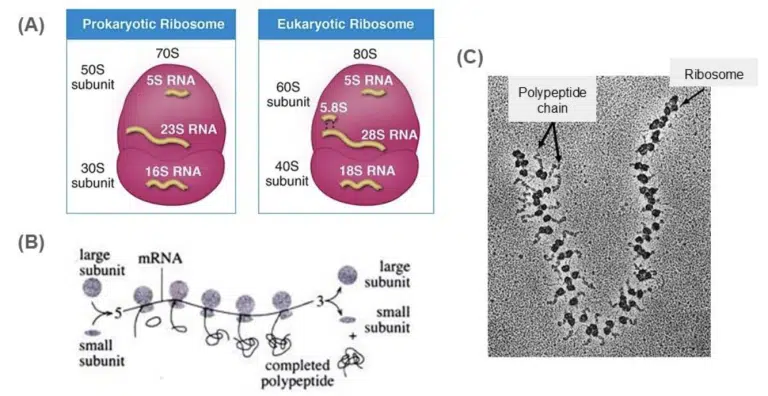
Fig 3. Schematic diagrams of a (A) DNA nucleotide and (B) RNA nucleotide.
Note that the deoxyribose sugar in DNA lacks the 2’ hydroxyl group (hence the name deoxyribose) compared to the ribose sugar in RNA. Also, instead of T, RNA uses U to undergo complementary base pairing with A. (C) A diagram of the DNA double helix structure. Notice the 5’ to 3’ directionality of each DNA strand (the phosphate group is facing the 5’ side!), and the hydrogen bonds between each base pair.
The human genome is approximately 3 billion base pairs long. For diploid (carrying 2 sets of chromosomes) creatures like us humans, that’ll be a total of 6 billion base pairs. Each base pair is approximately 0.34nm long, and hence the total length of all the DNA in a single cell would stretch up to 2m long! That’s way taller than this author! How does a microscopic cell fit all that genetic material into its nucleus? Just like your material belongings, some Marie Kondo organization is needed to ensure that genetic material is neatly packed away, but can be taken out of storage when it is needed. Here, we will dive into the levels of organization or “packing” of DNA.
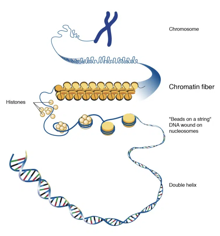
Fig 4. An illustration demonstrating the levels of DNA packing and organization.
At the most basic level of packing, DNA is wrapped around histone octamers to form a nucleosome. The nucleosomes can be further packed and compacted into chromatin fibers, which can then be packed into chromosomes.
At the most basic level of organization, DNA double helices are wrapped around a histone octamer like beads on a string. Each octamer consists of a pair of H2A, H2B, H3 and H4 histone proteins. Histones contain positively-charged lysine “tails” which stick out of the octamer and interact with negatively charged DNA, allowing the DNA strand to associate itself with the histone octamer. A complex between the DNA wrapped around its histone octamer is referred to as a nucleosome. We will later learn that many proteins can bind to histones to either “open” or “close” DNA for transcription and subsequent gene expression.
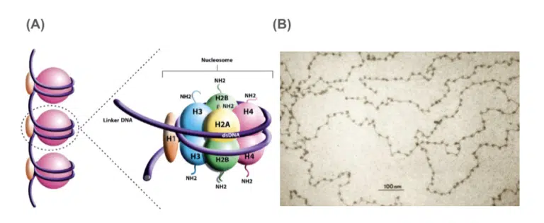
Fig 5. (A) A nucleosome consists of DNA wrapped around a histone octamer.
Each octomer consists of a pair of H2A, H2B, H3 and H4 proteins, and have positively charged lysine tails to interact with the negatively charged DNA. H1 histone helps to stabilise the nucleosome complex. (B) Transmission electron microscopy image of nucleosomes. Observe the “beads on a string” appearance of the nucleosomes.
Nucleosomes can be further packaged and compressed into chromatin fibers. Chromatin can be either loosely (euchromatin) or tightly packed (heterochromatin). Euchromatin can be observed as lighter regions within a nucleus and remains uncoiled and transcriptionally active. Heterochromatin on the other appears dark within a nucleus, as it is highly condensed and transcriptionally inactive. Association with other scaffold proteins can further promote condensation of chromatin.
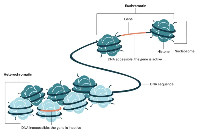
Fig 6. Illustration showing the difference in packing between euchromatin and heterochromatin.
Euchromatin is loosely packed, allowing the gene of interest to be accessible to transcription machinery. Hence, euchromatin is transcriptionally active. On the other hand, heterochromatin is tightly packed and stashes away the gene of interest, preventing its transcription and expression.
The most compact the DNA can go! To ensure accurate division of genetic material during cell division, DNA is packed into chromosomes with the help of condensin, a large protein complex that facilitates chromosomal assembly and subsequent segregation during cell division. Each chromosome has a centromere, which divides the chromosome into 2 sister chromatids. Humans have 2 sets of 22 chromosomes, and a pair of sex chromosomes, which bring the total number of chromosomes to 46. The tips of the chromosomes are capped with telomeres, which consist of telomeric DNA. As we will later learn in DNA replication, telomeric DNA are essential in protecting the genome against the end-replication problem, and maintain the integrity of the genome over successive rounds of cell division.
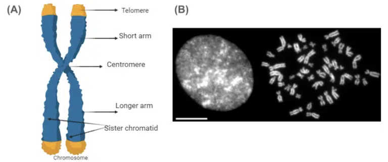
Fig 7. (A) Illustration of a chromosome at metaphase.
Chromosomes would only be this compact during cell division, which takes place after the S phase. By then, the cell would have replicated its DNA, and each chromatid on the chromosome is 1 copy of DNA. Each chromosome has a centromere which the mitotic spindle would attach to during mitosis. The tips of every chromatid is also capped with telomeres, which protect the integrity of the chromosome’s DNA over repeated rounds of DNA replication and cell division. (B) Fluorescence microscopy image of a cell’s nucleus outside of mitosis on the left, and the cell’s chromosomes at mitosis on the right. Here, the cell’s DNA has been labelled with fluorescent molecules to facilitate identifying and imaging. Notice how neatly compact the chromosomes are. Aren’t they much easier and better suited for splitting DNA evenly and accurately during cell division? We’ve made it to the end of this introductory article! So far, we have covered the basics of eukaryotic genomics. Be sure to be familiar with the contents of this article, as we will definitely be revisiting many of these concepts in future articles!
You might want to download a pdf copy of this article for future reference!
Click the white download button below, enter your email, and the pdf file will be delivered to your inbox! (Remember to check spam!)
The Science of Studying provides live online tuition via Zoom classes for Combined/Pure Chemistry, Biology, and Physics. To date, we have taught 800+ students over 12 years.
In case you are wondering, yes – there is a science behind studying!
At Science of Studying, we use our SOS system™ to teach our classes so that even last-minute students can see remarkable improvements in their grades – without mind-numbing memorisation of textbooks and without the drudgery of doing numerous assessment books.
All these conducted in a fun, interactive, stress-free online environment.
If you need help with your Chemistry, Biology, and Physics subjects, do reach out to us and we will see what we can do to help.
Contact Us: Click Here
Admin number: +65 88082348

The SOS system™️ guides students through an effective process of:
Join our proven online tuition programs and see real improvements in understanding, confidence, and school results.
Book a free trial lesson and start the journey today or discover more below:
A little more about ourselves…
The Science of Studying provides live online tuition via Zoom classes for Combined Chemistry, Combined Biology, Pure Chemistry, Pure Biology, JC Chemistry and JC Biology. To date, we have taught more than 800 students over 12 years.
In case you are wondering, yes – there is a science behind studying! At Science of Studying, we use our SOS system™️ to teach our classes so that even last-minute students can see remarkable improvements in their grades – without mind-numbing memorisation of textbooks and without the drudgery of doing numerous assessment books.
The SOS system™️ guides students through an effective process of:
All these conducted in a fun, interactive, stress-free online environment.
If you need help with your Chemistry and Biology subjects, do reach out to us and we will see what we can do to help.
Website: https://thescienceofstudying.com/
Admin number: +65 88082348
You might want to download a pdf copy of this article for future reference!
Click the blue download button, enter your email, and the pdf file will be delivered to your inbox! (Remember to check spam!)
WhatsApp us